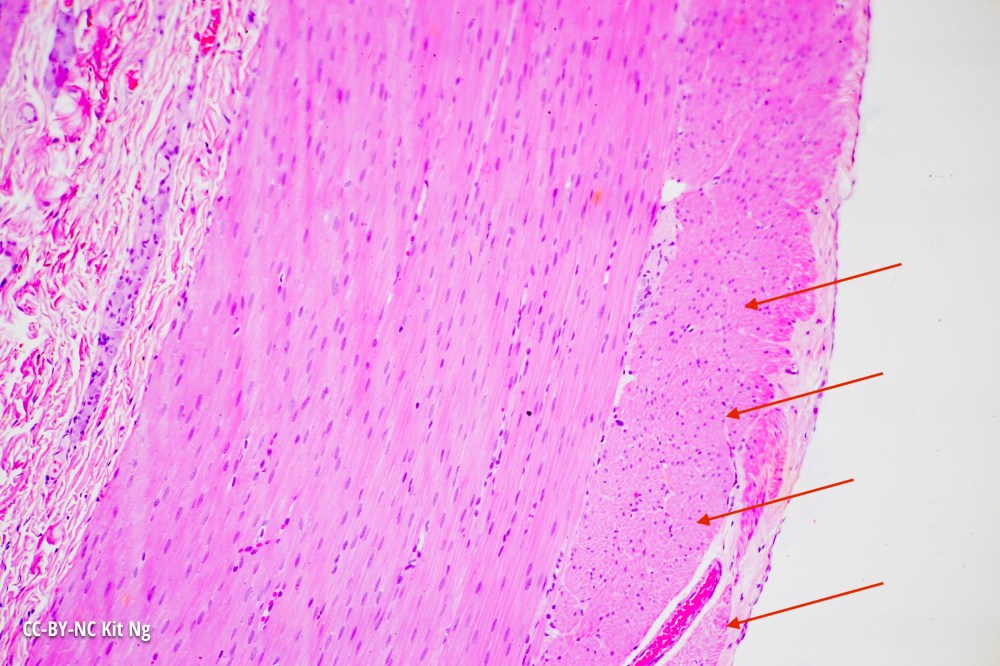
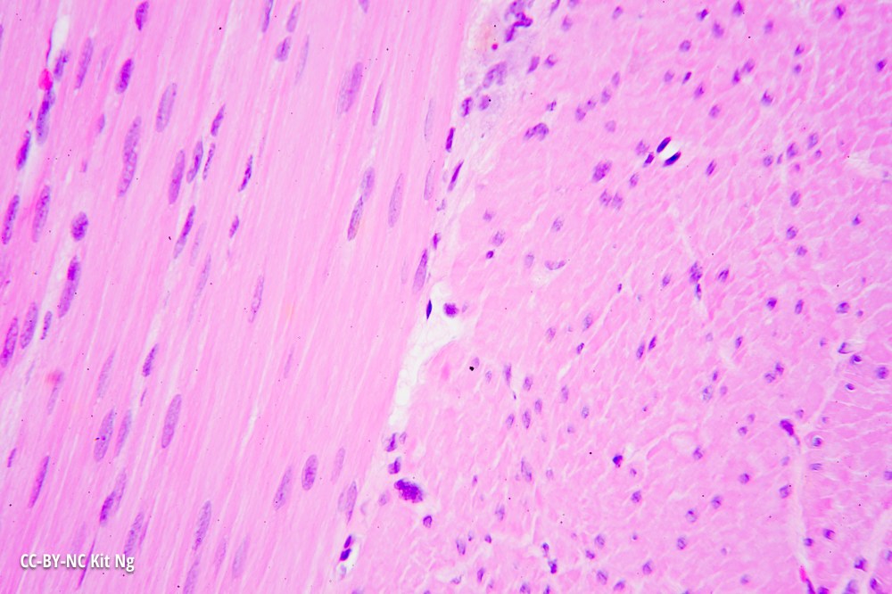
The large intestine is the penultimate portion of the digestive tract, between the small intestine and the rectum. The muscularis externa layer continues to separate into the circular and the longitudinal layer, with the exception that the longitudinal layer forming a muscular ribbon known as the teniae coli (at the pointers).
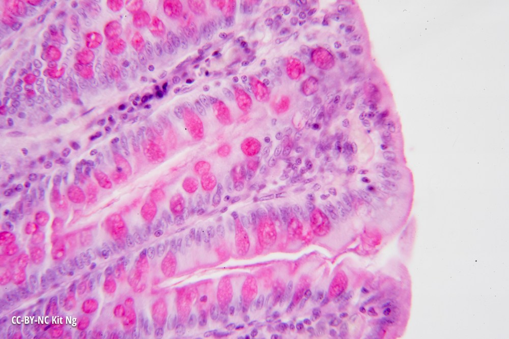
The large intestine absorbs water, electrolytes, and nutrients that are metabolized by the colonic bacteria, such as vitamin K. The remaining substance is mixed with mucus, produced by the numerous goblet cells in the colon, and becomes feces. The following picture stains specifically for mucus-producing cells.
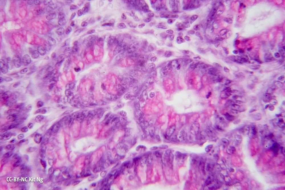
Finally, the simple columnar epithelial tissues are organized into intestinal (colonic) crypts with the lumen in the middle.