Fibrocartilage

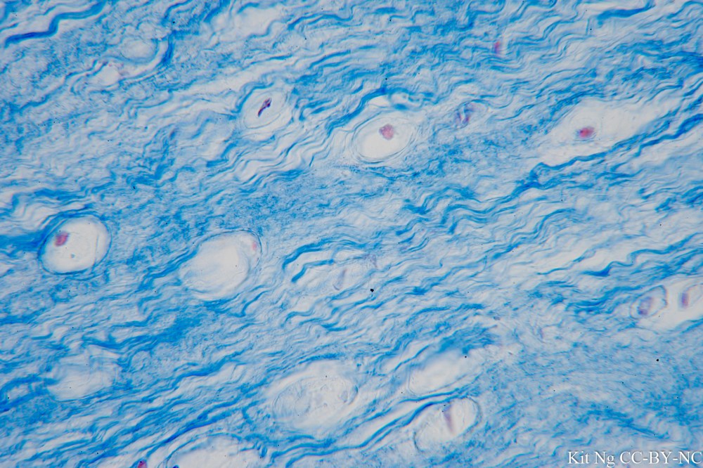
Fibrocartilage is similar to hyaline cartilage but with the addition of thick and dense collagen fibers that are deposited in the extracellular matrix.  It can be found in locations where shock absorption is needed such as intervertebral discs and symphyses joints.

_DSC4871.JPG
Fibrocartilage.  Lacunae with chondrocytes and the collagen fibers around can be seen.  (TM: 100x, picture taken with a Nikon Plan Apo 10/0.4 on Sony A7ii)
_DSC4873.JPG
The collagen fibers around the lacunae can be seen in this picture of Fibrocartilage.  (TM: 630x, picture taken with a Zeiss Planapo 63/1.4 Oil on Sony A7ii)
untitled shoot-2960.JPG
Another fibrocarilage specimen highlighting the collagen fibers.  (TM: 400x, picture taken with a Zeiss F40/0.8 on Sony A7ii)
_DSC5037.JPG
A different fibrocartilage specimen using a different staining method to stain for the collagen fibers.  (TM: 100x, picture taken with a Nikon Plan Apo 10/0.4 on Sony A7ii)
_DSC5038.JPG
Same specimen as above.  The chondrocytes in their respective lacunae could be seen in this view along with the collagen fibers around them.  (TM: 400x, picture taken with a Zeiss Planapo 40/1.0 Oil on Sony A7ii)

Leave a comment