Skeletal muscle tissue is easily distinguished by the striation (thus also known as striated muscle). They are arranged as long cylinders and since fused from development, contain multiple nuclei within each muscle fiber/cell.
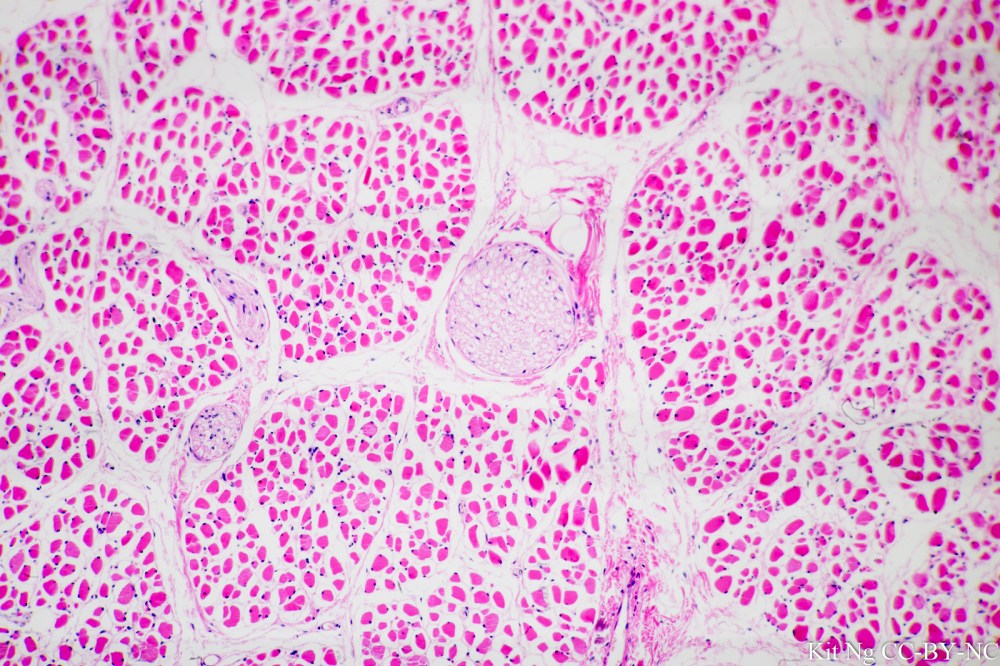
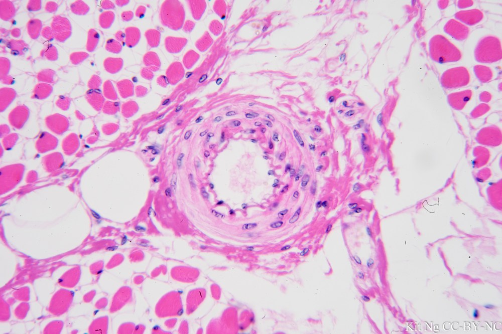
Muscle, transverse sectioned. The endomysium surrounding each muscle fiber, perimysium surrounding each fascicle and peripheral nerve can be seen. (TM: 100x, picture taken with a Nikon Planapo 10/0.4 on Sony A7ii)Muscle, t.s. The muscle fiber and an accompanying arteriole could be seen. (TM: 400x, picture taken with a Zeiss Planpo 40/1.0 Oil on Sony A7ii)Muscle, longitudinal sectioned. The cylinder shape muscle fiber and multiple nuclei can be seen. (TM: 400x, picture taken with a Zeiss Planapo 40/1.0 Oil on Sony A7ii)Muscle, l.s., with emphasis on showing the striations. Annotated version here. (TM: 630x, picture taken with a Zeiss Planapo 63x/1.4 Oil on Sony A7ii)Muscle, l.s., with emphasis on showing the striations. (From a different slide than above. TM: 1000x, picture taken with a Neofluar 100/1.3 Oil on Sony A7ii)