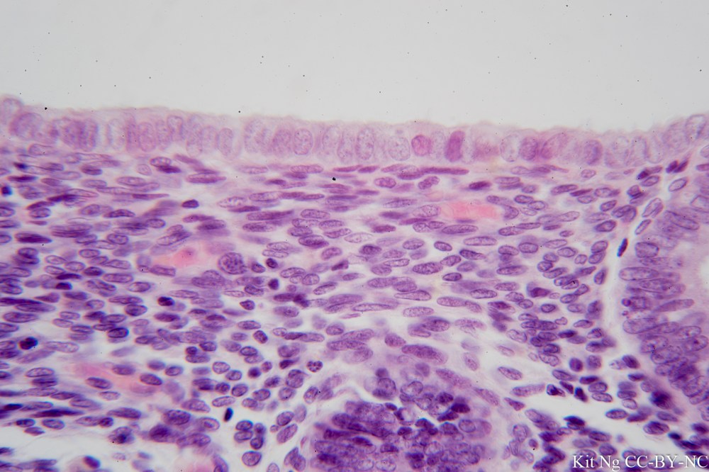
Simple columnar epithelium contains cells that are rectangle in shape and have their nuclei arranged on the basal side. Their nuclei also tend to look oval shape. This type of epithelium can be found in the digestive tract, uterine tube and central canal of the spinal cord.
In the digestive tract, structures called microvilli (or Brush border) could be found on the apical side of the epithelium. These structures increase the surface area for more efficient absorption. Goblet cells that secret mucus could also be found.
In the uterine tube, cilia could be found on the apical side. Their function is to propel the egg cell towards the uterus, failure of which could lead to ectopic or tubal pregnancy.
Simple columnar epithelium of the small intestine. Notice the goblet cells (the cells that contain a white spot) and the fuzzy brush border on the apical side. Annotated version here.(TM: 400x, picture taken with a Zeiss F40/0.8 on Sony NEX-6)Simple columnar epithelium showing the microvilli/brush border. (TM: 1000x, picture taken with a Zeiss Neofluar 100/1.3 on Sony NEX-6)Simple columnar epithelium of the gall bladder. (TM: 400x, picture taken with a Zeiss Planapo 40/1.0 Oil on Sony A7ii)Simple columnar epithelium of the duodenum. (TM: 400x, picture taken with Zeiss Planapo 40/1.0 Oil on Sony A7ii)Simple columnar epithelium of the Human uterus in follicular phase. (TM: 630x, picture taken with a Zeiss Planapo 63/1.4 Oil on Sony A7ii)