Jejunum and ileum comprises the distal portion of the small intestine after the duodenum. Though the basic layers (mucosa, submucosa, muscular is propria and adventia/serosa) remain the same and the mucosa also form villi, there are a few structural changes that set the duodenum and jejunum/ileum apart:
Absence of Brunner’s Gland
Villi becoming progressively shorter
Increasing amount of goblet cells in the epithelium
Mucosa forming circular fold known as plicae circulares
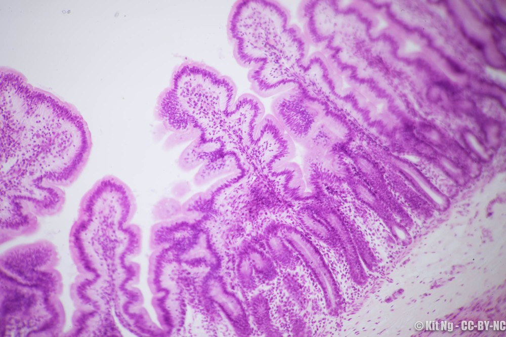
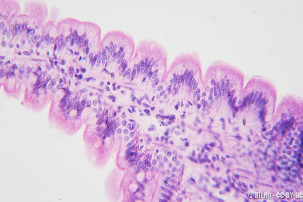
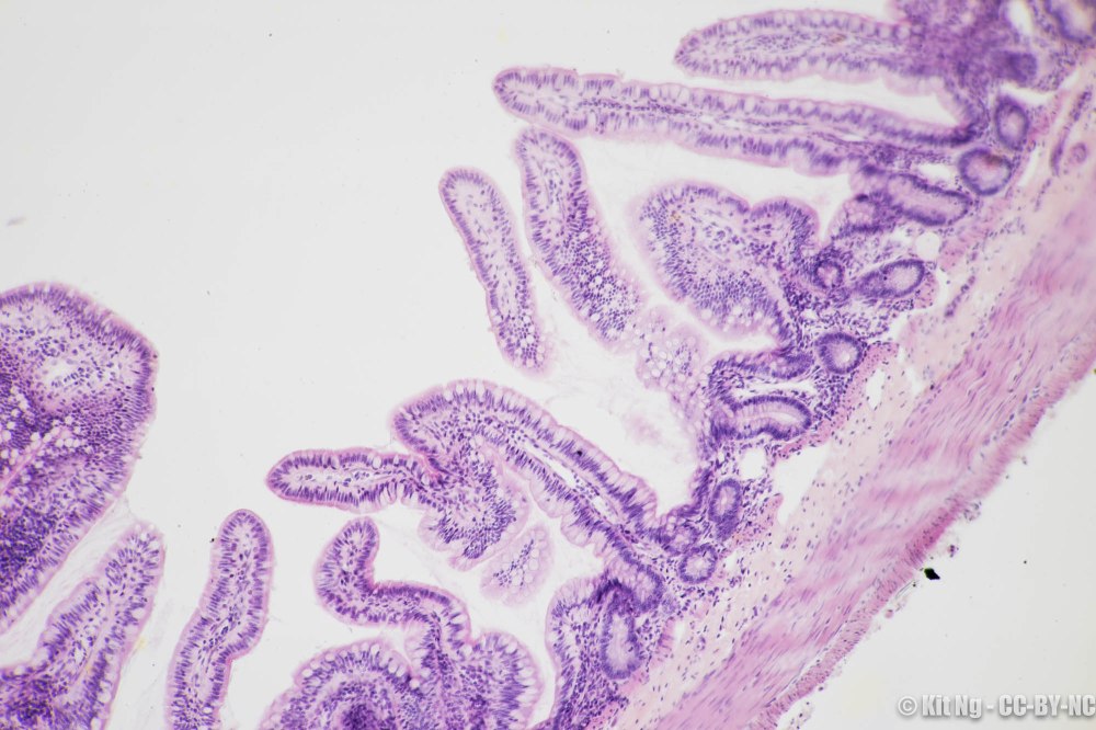
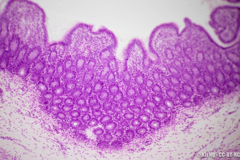
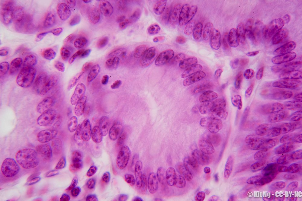
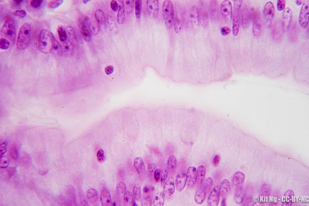
Mucosa of the Jejunum, showing the circular fold (plicae circulares) along the length of the villi. (TM: 100x, picture taken with a Nikon Planapo 10/0.4 on Sony A7ii)Another specimen of the villi of the Jejunum, showing the circularly arranged fold known as plicae circulares along the villi. (TM: 400x, picture taken with a Zeiss Planapo 40/1.0 Oil on Sony A7ii)Villi of the Ileum. Notice the presence of numerous goblet cells. Plicae circulares not prominent in this view. (TM: 100x, picture taken with a Nikon Planapo 10/0.4 on Sony A7ii)Villi of the Ileum showing the simple columnar epithelium and goblet cells. (TM: 630x, picture taken with a Zeiss Planapo 63/1.4 Oil on Sony A7ii)Another specimen of the Ileum showing the shorter villi and the Crypts of Lieberkuhn in the mucosa. These contain stem cells that regenerate the overlying epithelium. (TM: 100x, picture taken with Nikon Planapo 10/0.4 on Sony A7ii)Crypt of Lieberkuhn found in Jejunum. Notice the mitotic cell in the upper left corner of this crypt. (TM: 1000x, picture taken with a Zeiss Planapo 100/1.3 Oil on Sony A7ii)Intraepithelial lymphocytes found in the villi of the Ileum. (TM: 1000x, picture taken with a Zeiss Planapo 100/1.3 Oil on Sony A7ii)