Neurons as a cell type have very diverse structures. They could be classified by their modalities and morphologies. Their functions are to conduct messages to and from the Central Nervous System and Peripheral Nervous System, or within each system.\
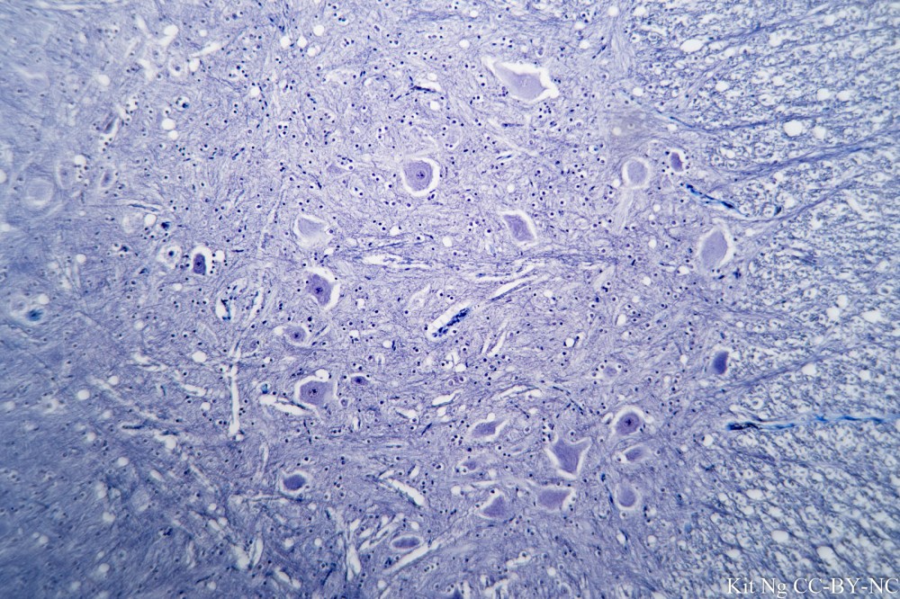
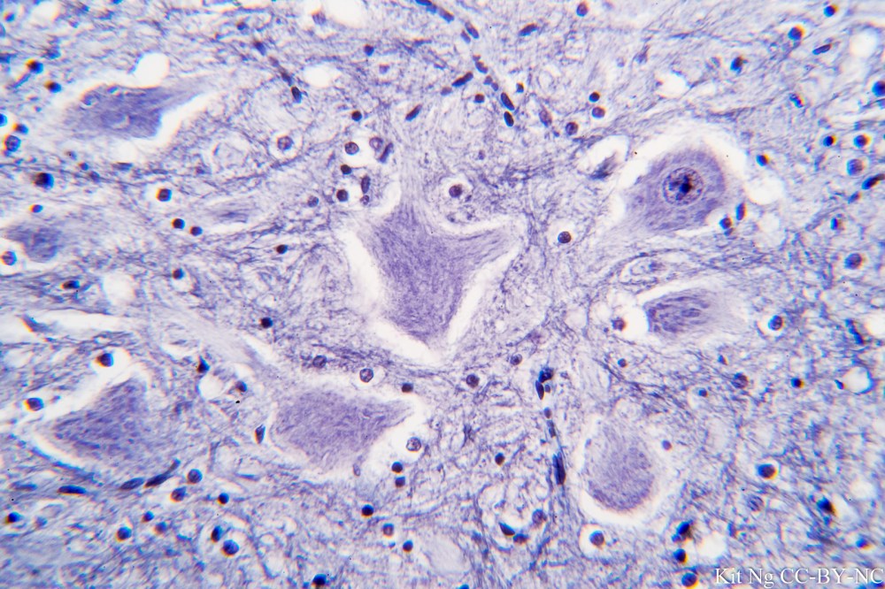
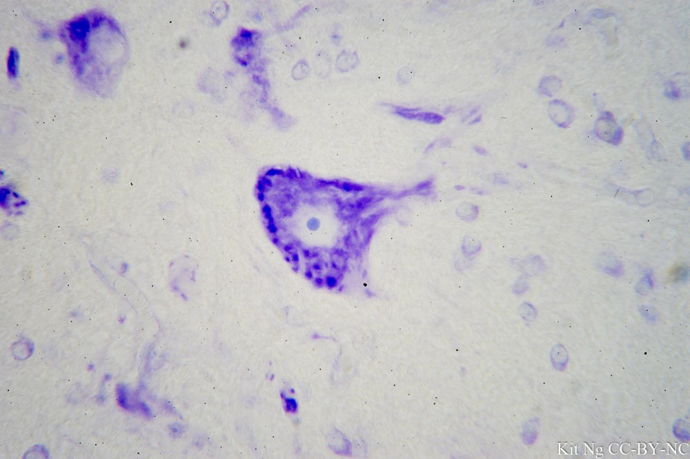
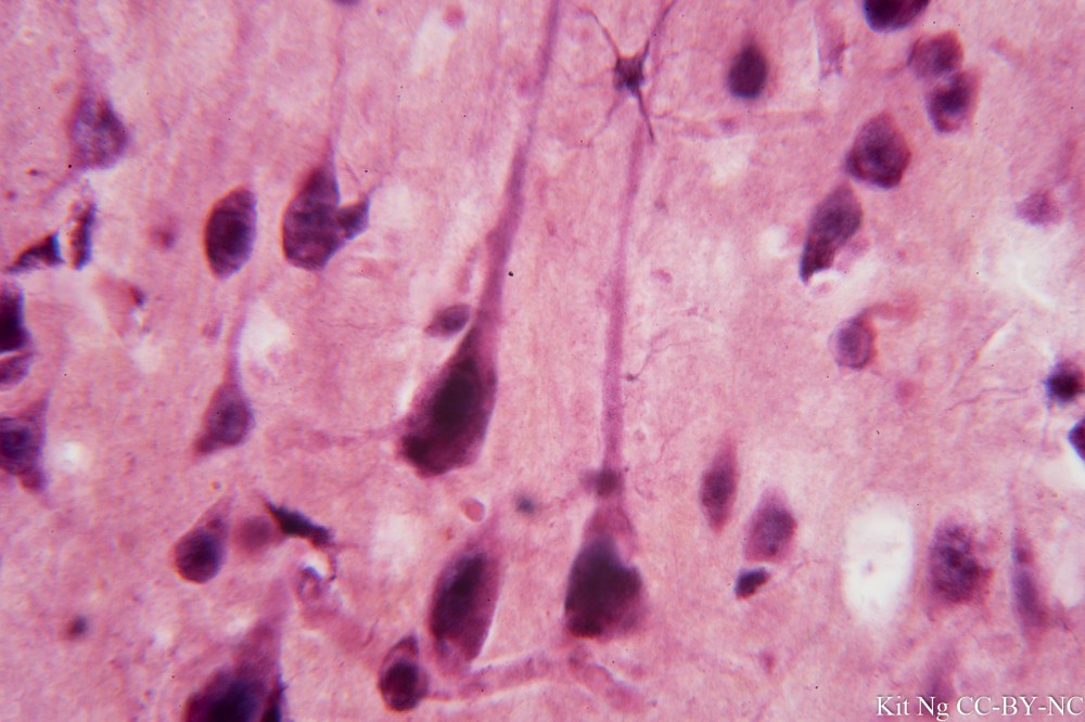
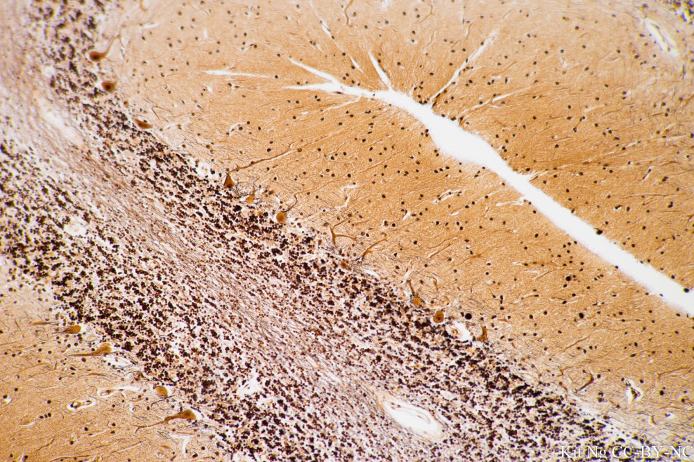
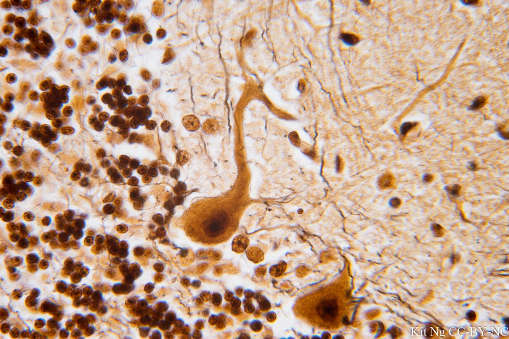
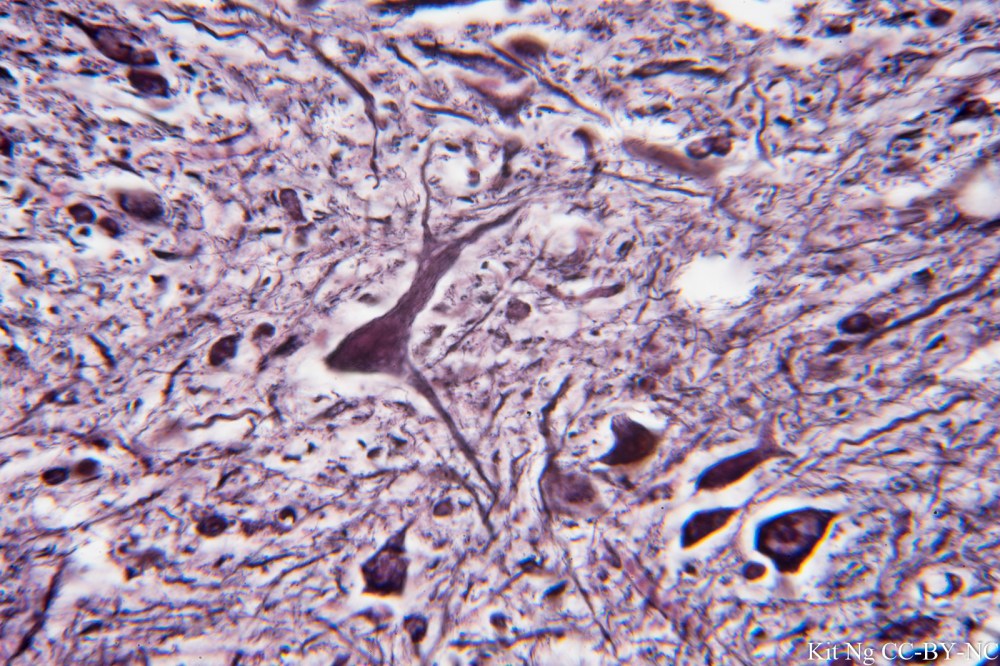
Giant motorneuron of the Spinal Cord, trichrome stain. (TM: 100x, picture taken with an Olympus DPlan 10/0.25 on Sony A7ii)Giant motorneuron of the Spinal Cord, trichrome stain. (TM: 400x, picture taken with a Zeiss F40/0.8 on Sony A7ii)Nissl bodies of a motorneuron stained with Nissl stain. These are rough endoplasmic reticulum and site of protein synthesis. Annotated version here. (TM: 630x, picture taken with a Zeiss Planapo 63/1.4 Oil on Sony A7ii)Giant pyramidal cells of the Cerebral Cortex. (TM: 630x, picture taken with a Zeiss Planapo 63/1.4 Oil on Sony A7ii)Cerebellum with silver impregnation. The layer of Purkinje cells at the border of the molecular layer and granular cell layer can be seen. (TM: 100x, picture taken with a Nikon Plan Apo 10/0.4 on Sony A7ii)Purkinje cell with dendrites extending into the outer molecular layer and the fine axon into the granular cell layer that is hard to observe. Annotated version here. (TM: 630x, picture taken with a Zeiss Planapo 63/1.4 Oil on Sony A7ii)Spinal Cord staining for the neuro-fibril. (TM: 630x, picture taken with a Zeiss Planapo 63/1.4 Oil on Sony A7ii)