Skin Layers

There are four layers of what one consider to be skin, from superficial to deep, they are epidermis, dermis, hypodermis, and deep fascia.

160810_untitled_4888.JPG
Skin Layers. (TM: 100x, picture taken with a Nikon Plan Apo 10/0.4 on Sony A7ii)

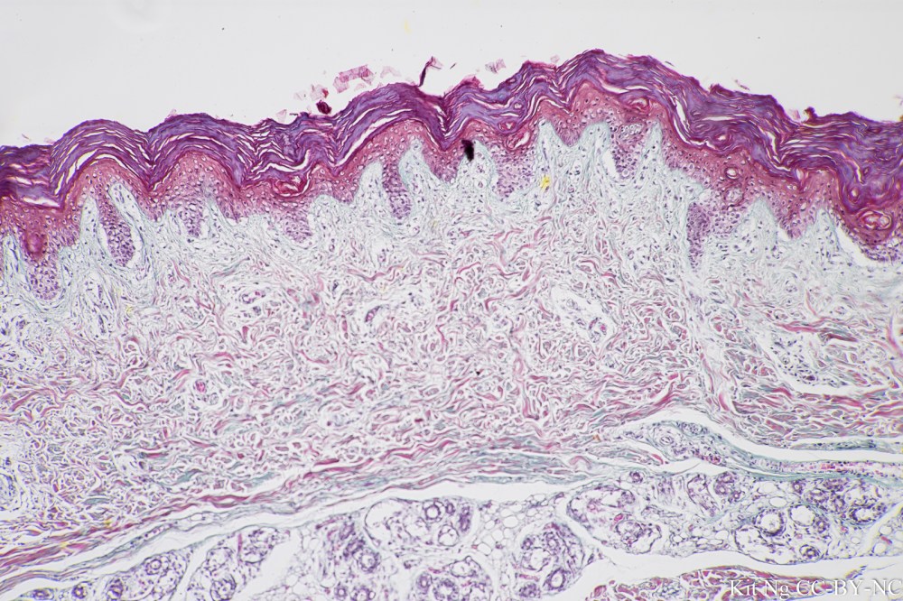
Epidermis is formed by stratified squamous epithelial tissue, keratinized type:

_DSC4936
Stratified squamous epithelial tissue.  Annotated version can be found here.  (TM: 100x, picture taken with a Nikon Plan Apo 10/0.4 on Sony A7ii)

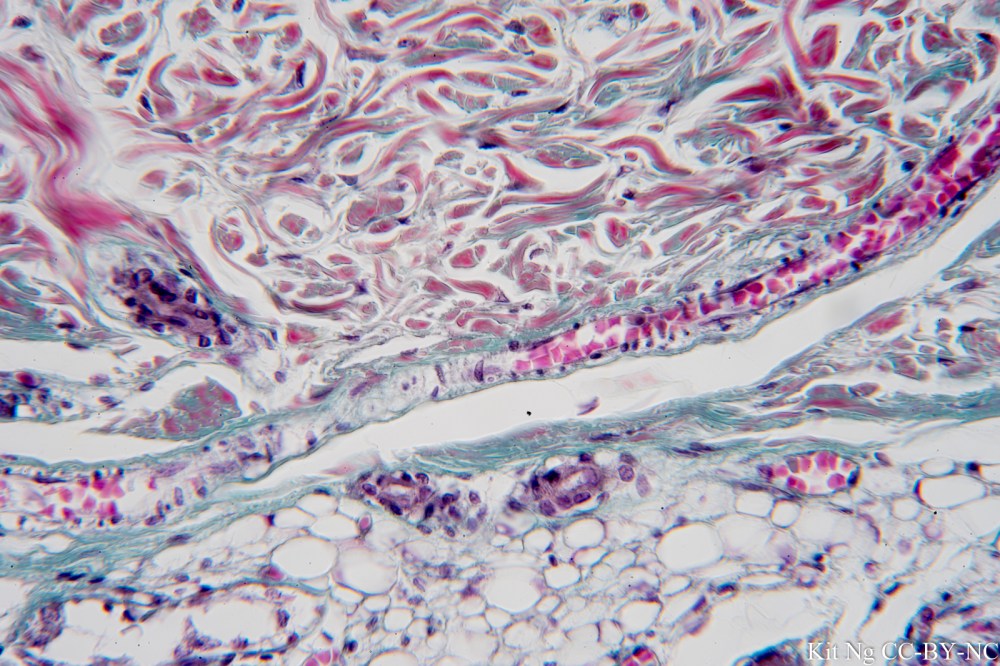
Dermis has two layers: papillary dermis and reticular dermis.  The deeper reticular dermis is composed of mostly dense irregular connective tissue that makes it resistant to pulling forces in many directions.  Structures that can be found in the dermis are: Messiner’s corpuscle, Pacinian corpuscle, hair follicle, arrector pili, sebaceous (oil) gland, and sweat gland.

_DSC4938
Dermis showing the dense irregular connective tissue, hair follicle, oil gland, and sweat gland.  Annotated version can be found here. (TM: 100x, picture taken with a Nikon Plan Apo 10/0.4 on Sony A7ii)

Hypodermis is composed of mostly adipose tissue.  It is also known as the subcutaneous layer and superficial fascia.

160810_untitled_4889.JPG
Dermis/Hypodermis junction showing collagen fibers of the dense irregular CT and adipose tissue.  (TM: 400x, picture taken with a Zeiss Planapo 40/1.0 Oil on Sony A7ii)

Under the hypodermis would be the deep fascia that wraps around the skeletal muscle.

Leave a Reply

Fill in your details below or click an icon to log in:

WordPress.com Logo

You are commenting using your WordPress.com account. Log Out /  Change )

Facebook photo

You are commenting using your Facebook account. Log Out /  Change )

Connecting to %s